
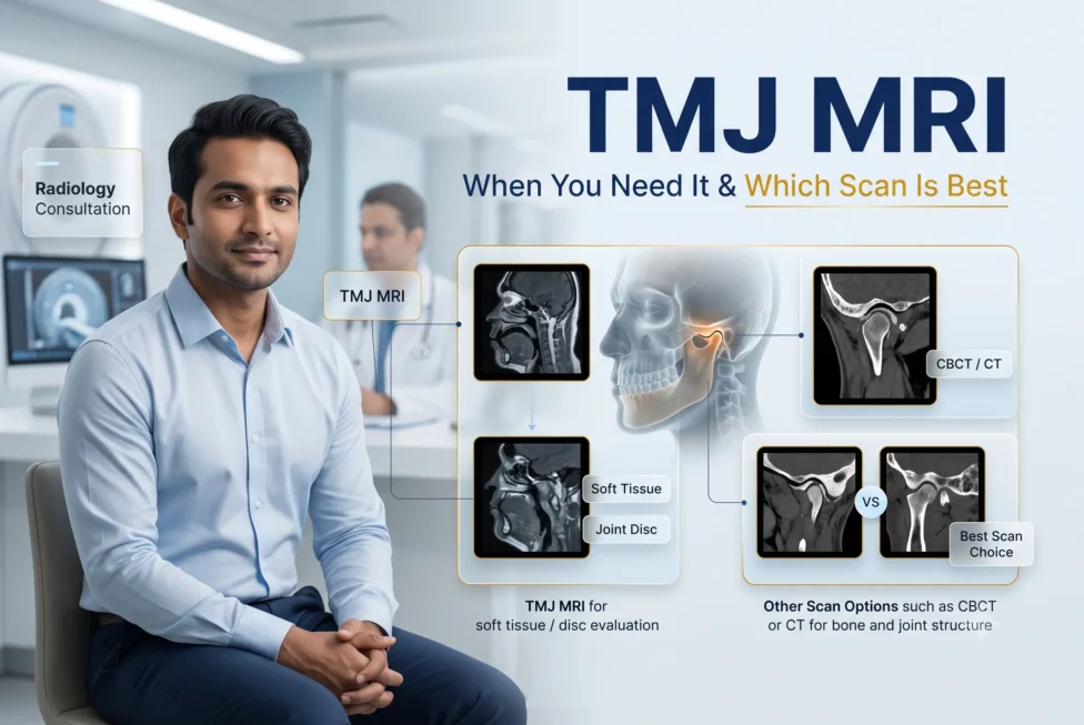
Most people with TMJ pain do not automatically need an MRI. Diagnosis usually starts with the symptom history and a clinical examination. MRI is most useful when a clinician needs to assess the TMJ disc, joint fluid or other soft tissues. CBCT is better for bone, while a panoramic X-ray is a limited 2-D overview. The right scan is the one that answers a specific question and could change care.
You may have jaw pain, clicking or limited opening and wonder whether a scan will finally show the cause. The problem is that ‘a TMJ scan’ is not one test. MRI, CBCT, medical CT and panoramic X-ray show different anatomy, and ordering the wrong one can add cost without answering the question that matters.
| The most useful rule
Clinical question first, scan second. The 2026 ADA–AAOMR recommendations say imaging should follow the medical and dental history and clinical examination, and should not be routine in TMD diagnosis. Ask what the clinician expects the image to show and how that result could change treatment or referral. |
When is TMJ imaging not usually the first step?
Imaging may not be needed at the first visit when symptoms and examination fit a common TMD pattern, there is no trauma or progressive change, and the initial plan would be conservative regardless of the image. NIDCR notes that TMD diagnosis begins with a detailed history and examination; imaging may be added when needed.
Painless clicking alone is common and generally does not require treatment. Even when pain is present, a clinician may first check the jaw muscles, joint tenderness, range and pattern of movement, bite change, teeth, ears, head and neck, and relevant medical history. Imaging becomes more useful when that assessment raises a focused question.
| Imaging sooner may be appropriate when
There is facial trauma or suspected fracture/dislocation; persistent function-limiting locking or restricted opening; progressive bite or facial change; suspected arthritis or structural bone disease; significant swelling, fever or a mass; neurological symptoms; or pain that does not fit a typical dental or TMD pattern. |
TMJ MRI vs CBCT vs X-ray: what does each show?
| Question | TMJ MRI | CBCT / CT | Panoramic X-ray |
| Best at | Soft tissue: disc position and shape, joint fluid, marrow and inflammatory change | Bone: cortical detail, erosions, osteophytes, resorption, developmental change and fracture | A broad 2-D overview of the jaws, teeth and gross bony abnormality |
| Does not answer well | Fine bony surface detail compared with CBCT or CT | CBCT does not show the TMJ disc reliably; medical CT has limited disc detail | Disc position, joint fluid, early soft-tissue change or fine 3-D bone detail |
| Radiation | No ionising radiation; MRI safety screening is still required | Uses ionising radiation; protocol and field of view should match the clinical question | Uses a small amount of ionising radiation |
| Common role | Function-limiting suspected internal derangement or soft-tissue disease | Suspected bony disease, progressive structural change or trauma | Possible initial screen, but low sensitivity limits definitive TMJ diagnosis |
Medical CT and CBCT both provide strong bony detail. Medical CT can also show surrounding soft-tissue spaces better than CBCT and may be chosen in acute or complex trauma. For a dedicated disc question, however, MRI remains the reference imaging method.
Which TMJ scan matches which clinical question?
- Is the disc displaced or not moving normally? A dedicated TMJ MRI is the appropriate soft-tissue study when the suspected internal derangement is functionally limiting and confirmation could change management.
- Is there joint fluid, inflammation or marrow change? MRI can show joint effusion and soft-tissue or marrow findings. The radiologist and referring clinician decide whether contrast is necessary; it is not automatic for every TMJ MRI.
- Is the concern bone erosion, osteoarthritis, resorption or developmental change? CBCT is generally preferred for detailed bony assessment.
- Was there a blow to the jaw or suspected fracture? CBCT or medical CT may be selected for hard-tissue injury. Urgent trauma care comes before elective imaging decisions.
- Could the pain come from teeth or another jaw condition? A panoramic or targeted dental X-ray may help screen the teeth and jaws, but it cannot show the TMJ disc.
What does a normal TMJ MRI mean?
A normal MRI means the radiologist did not identify a significant abnormality in the structures and sequences examined. It does not mean the pain is imaginary. Muscle-related TMD, referred pain, headache disorders, neuropathic pain and dental or ear conditions may not be explained by a TMJ disc image.
The reverse is also important: an imaging finding is not automatically the source of pain. The AAOMR–AAOP position statement notes that TMJ pain, function and disability do not necessarily correlate with radiologic disc-displacement findings. The report must therefore be interpreted alongside the examination and the patient’s main problem.
If imaging is normal but symptoms persist, the next step is not necessarily a more expensive scan. The clinician may revisit the diagnosis, look for muscle or non-TMJ causes, review whether the correct area and protocol were imaged, or coordinate with another medical or dental specialty.
What should you expect during a TMJ MRI?
MRI uses a strong magnetic field and radio waves rather than ionising radiation. A TMJ examination may include images with the mouth closed and open so the disc and joint can be assessed in different positions. You must keep still, and the scanner can be noisy.
- Tell the imaging team about implanted devices, metal in the body, previous surgery, pregnancy, and any history of metal injury.
- Mention claustrophobia or anxiety before the appointment so the facility can discuss suitable options.
- Bring the referral, previous images and reports, and a current list of relevant medical conditions and devices.
- Ask whether the appointment is for a dedicated TMJ protocol rather than a general head or brain MRI.
Do not assume that contrast is required. The protocol depends on the diagnostic question, and the radiologist should decide when contrast adds useful information.
Five questions to ask before paying for a TMJ scan
| Question to ask | Why it matters |
| What exact diagnosis are we investigating? | A disc question, bone question and fracture question require different imaging. |
| How could the result change my treatment? | A scan is most useful when a possible result would alter management, referral or planning. |
| Have my previous images been reviewed? | Existing dental or medical images may answer part of the question and reduce unnecessary repeat exposure. |
| Is this a dedicated TMJ protocol? | Disc assessment commonly needs appropriate TMJ sequences and open- and closed-mouth views. |
| Who will interpret it and explain it with my examination? | The report should be matched to symptoms, function and clinical findings—not read in isolation. |
These questions are not about refusing useful imaging. They help make the investigation more precise. A well-selected scan can clarify diagnosis or treatment planning; a poorly selected scan may produce incidental findings or leave the original question unanswered.
TMJ assessment in Noida: choose the question before the machine
Airway TMJ & Sleep Center can assess jaw pain, locking, clicking, limited opening and bite change, then discuss whether imaging is likely to add useful information. The clinic should not be assumed to provide every imaging modality in-house; when a scan is indicated, the appropriate study can be coordinated with a qualified imaging centre and interpreted in the context of the examination.
Review the TMJ symptoms and warning signs guide, learn about the clinic’s TMJ pain assessment service, or contact the clinic to arrange an evaluation in Noida.
| Primary next step
Book a clinical TMJ evaluation before choosing a scan. Call +91 87566 15325. Airway TMJ & Sleep Center, C-98/10, C Block Market, Sector 41, Noida 201301. |
The bottom line
MRI is not automatically the best first test for every person with TMJ pain. It is the strongest option for disc and soft-tissue questions; CBCT is stronger for bone; a panoramic X-ray is a limited overview. The best imaging decision starts with a clinical examination, a precise diagnostic question and a plan for how the result will change care.