
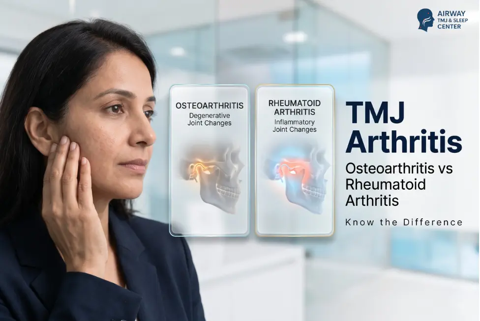
TMJ arthritis means an arthritic process affecting the jaw joint. It may be degenerative, as in osteoarthritis, or inflammatory, as in rheumatoid arthritis or another rheumatic disease. Both can cause pain near the ear, stiffness, grating, limited opening or bite change. Symptoms alone cannot identify the subtype: examination comes first, CBCT or CT answers bony questions, and MRI evaluates soft tissue, fluid and inflammation when indicated.
Jaw pain in a person with arthritis should not automatically be labelled clenching or osteoarthritis. The useful question is whether it reflects active inflammation, structural change, another pain source—or a combination.
What does arthritis mean in the TMJ?
The temporomandibular joints (TMJs) are the two moving joints in front of the ears. ‘TMD’ is the broader name for disorders of these joints, chewing muscles and related structures; arthritis is only one possible joint diagnosis.
Osteoarthritis involves gradual change in cartilage, bone and other joint tissues; it is not simply a joint being ‘worn out.’ Inflammatory arthritis is immune-driven. Rheumatoid arthritis can affect the jaw, as can other rheumatic diseases.
The categories can overlap. Long-standing inflammation may leave degenerative bone change, while muscle or disc pain may coexist. A scan description such as ‘osteoarthritic change’ does not explain every symptom or identify the original cause.
TMJ osteoarthritis vs inflammatory arthritis
| Clue | TMJ osteoarthritis pattern | Inflammatory arthritis pattern |
| Underlying process | Gradual structural change in cartilage, bone and other joint tissues. It is more complex than simple ‘wear and tear.’ | Immune-driven inflammation of the joint lining, as in rheumatoid arthritis or another rheumatic disease; it may later cause structural change. |
| Typical context | May follow age-related change, prior injury, altered joint loading or another joint process; sometimes no single cause is found. | Known or suspected systemic inflammatory disease, although jaw symptoms may occasionally prompt the wider evaluation. |
| Symptom pattern | Pain often changes with use; stiffness, crepitus and reduced movement may develop gradually. | Pain or stiffness may occur at rest and with use; prolonged morning stiffness, flares or rapid functional change raise concern. |
| Clues outside the jaw | May occur without systemic inflammatory symptoms, although osteoarthritis can affect other joints too. | Swollen or warm joints, prolonged morning stiffness, fatigue, low-grade fever, psoriasis or other disease-specific features may coexist. |
| Imaging question | Are there bony degenerative or remodelling changes, and are they stable or progressive? | Is there active soft-tissue inflammation or effusion, bony damage, or both—and does it fit the systemic picture? |
| Care team | Dentist/TMJ or orofacial-pain clinician; radiology and maxillofacial input when indicated. | Dental/TMJ clinician plus physician or rheumatologist; paediatric team for juvenile idiopathic arthritis. |
| Important limit | Bone changes do not prove that every symptom comes from arthritis. | Jaw symptoms alone do not diagnose rheumatoid or another systemic arthritis. |
Symptoms that deserve a closer look
Local symptoms may include pain in front of the ear, stiffness, reduced opening, painful grating, chewing difficulty, locking or bite change. A painless click alone is common and does not prove arthritis.
Inflammatory clues include stiffness lasting more than 30 minutes after waking, swollen joints elsewhere, fatigue, low-grade fever or known inflammatory disease. These support medical review; they do not allow a jaw-only rheumatoid arthritis diagnosis.
In a Finnish sample of 6,331 adults, 64% of participants with seronegative RA and 60% with seropositive RA had at least one clinical TMD sign. These were signs—not complete TMJ diagnoses—and age and other factors also mattered.
| Do not wait for a routine appointment
Emergency now: trouble breathing or swallowing, rapidly spreading face or neck swelling, serious trauma, severe bleeding, collapse/confusion, or a jaw stuck open and unable to close. Do not force the jaw into position. Same-day urgent assessment: fever with new swelling, warmth or redness near the joint; sudden severe pain with rapid loss of opening; pus or a suspected dental infection; or marked deterioration in someone who is immunosuppressed. Rare TMJ infection can resemble arthritis. Prompt specialist review: a progressively changing bite or new open bite, increasing facial asymmetry, rapid loss of function, unexplained systemic inflammatory symptoms, or jaw change in a child or adolescent with possible juvenile idiopathic arthritis. |
Which scan is used for TMJ arthritis?
There is no single ‘best scan’ for every person. The clinician first decides what needs to be learned and whether the result would change care. The 2026 ADA–AAOMR recommendations specifically advise against routine imaging before a clinical assessment.
| Assessment | Best at showing | What patients should know |
| Clinical examination | Pain location, familiar pain, movement, bite, sounds, muscles and competing dental or medical causes. | This comes first. Imaging is selected only when it can answer a question that affects diagnosis or care. |
| Panoramic X-ray | Broad dental and jaw overview; gross bony abnormality. | Useful in some initial pathways, but it can miss or understate detailed TMJ bone change. |
| CBCT / CT | Bone surfaces and changes such as erosion, flattening, sclerosis, osteophytes, remodelling or resorption. | The 2026 ADA–AAOMR recommendations prefer CBCT for bony TMJ components, including degenerative disease and systemic arthritides. It uses ionising radiation and does not replace MRI for soft tissue. |
| MRI | Disc and other soft tissue, joint fluid/effusion and inflammatory change; also bone-marrow information. | MRI is the reference standard for soft-tissue TMJ pathology. It has no ionising radiation, but it is not automatically needed for every jaw-pain visit. |
| Medical tests | The wider inflammatory pattern, guided by a physician or rheumatologist. | No single blood test proves that jaw pain is TMJ arthritis. Results are interpreted with symptoms, examination and systemic history. |
Why the report must be matched to the person
Imaging and symptoms do not always match. Bone remodelling may occur with little pain, while inflammation or pain may precede dramatic bony change. Ask which finding matches the examination, whether older images show progression and what decision the scan changes.
How is TMJ arthritis treated?
Treatment is cause-based: stable degeneration, active inflammation, disc or muscle pain and infection require different pathways. The first aim is to protect function while clarifying the diagnosis.
- Reduce avoidable overload temporarily. Choose easier-to-chew food during a flare, avoid gum and extreme opening, and stop repeatedly testing the joint.
- Use guided rehabilitation. A clinician or physiotherapist may select gentle movement and function goals. Aggressive stretching is not suitable for every inflamed, unstable or acutely infected joint.
- Review appliances and habits. An appliance is not an arthritis diagnosis and must be individually indicated and monitored for symptoms and bite change.
- Coordinate systemic disease control. A rheumatologist or physician leads rheumatoid or other inflammatory-disease medication decisions. Do not start, stop or change prescribed medicine because of this article.
- Escalate selectively. Injections, lavage or surgery are not one-size-fits-all first steps. They may be discussed when a clear diagnosis, severity and response to appropriate conservative or medical care justify them.
Evidence for local procedures remains limited. A 2025 review found only five eligible, varied and low-quality studies, so it could not draw firm efficacy conclusions.
Clinician-coordination checklist
| Team member | Information to coordinate |
| Dentist / TMJ or orofacial-pain clinician | Pain map, opening and movement, joint and muscle findings, bite change, dental causes, functional goals and the precise imaging question. |
| Physician / rheumatologist | Other joints and systemic symptoms, established diagnosis, disease activity, laboratory context and all systemic medication decisions. |
| Oral and maxillofacial radiologist | The most appropriate modality and protocol, a complete report, and comparison with prior imaging to distinguish stable remodelling from progression. |
| Physiotherapist / surgeon when indicated | Safe restoration of function after diagnosis, or specialist review when there is major structural damage, rapid loss of function or failure of appropriate conservative care. |
Bring these details to the appointment
- A timeline: first symptom, flares, morning pattern, triggers and change in mouth opening or bite.
- Other painful, swollen or stiff joints and any fatigue, fever, skin, eye or bowel symptoms.
- Your rheumatology diagnosis, medicine list and recent reports—without changing treatment yourself.
- Previous TMJ images/reports, appliance history, trauma or surgery details.
- A practical goal: eating, yawning, speaking, brushing or sleeping with less pain or restriction.
A special note for children and adolescents
TMJ arthritis in juvenile idiopathic arthritis can matter even when jaw pain is mild or absent because the joint contributes to facial growth and daily function. Jaw asymmetry, a changing bite, reduced opening or known JIA deserves coordinated paediatric rheumatology and dental/maxillofacial assessment rather than an adult self-care plan.
Questions to ask before accepting a treatment plan
- What evidence suggests arthritis rather than muscle, disc, dental or referred pain?
- Does the pattern look degenerative, actively inflammatory or mixed—and what remains uncertain?
- What exact question will an X-ray, CBCT/CT or MRI answer?
- Can the new scan be compared with an older one to check progression?
- Which clinician is leading systemic disease care, jaw function and imaging follow-up?
- What measurable change should trigger earlier review or a surgical opinion?
Coordinated TMJ arthritis assessment in Noida
Airway TMJ & Sleep Center can evaluate jaw-joint and muscle findings, document opening and bite changes, review available imaging and coordinate with a physician or rheumatologist when systemic arthritis is present. The aim is to answer the diagnosis and imaging questions before choosing a local procedure.
Review the TMJ symptoms and warning signs guide, see the jaw swelling location guide, or contact the clinic for a coordinated evaluation.
| Bring your medical and dental records together
Call +91 87566 15325. Airway TMJ & Sleep Center, C-98/10, C Block Market, Sector 41, Noida 201301. Fever with swelling, rapid deterioration or emergency symptoms need urgent medical or hospital assessment—not a routine TMJ appointment. |
The bottom line
TMJ arthritis is not one condition. Osteoarthritis is a degenerative structural process; rheumatoid and other inflammatory arthritides are immune-driven. Their pain, stiffness, crepitus, restriction and bite changes can overlap.
Start with history and examination, select imaging for a specific question, and connect jaw findings to the wider medical picture. A useful plan protects function, tracks change and gives each clinician a clear role.
| Medical disclaimer
This article is for education and cannot diagnose TMJ arthritis, rheumatoid arthritis, infection or another cause of jaw pain. It does not provide medication advice. Do not change prescribed rheumatology, dental or pain treatment without the treating clinician. Seek urgent or emergency care for the warning signs above. |